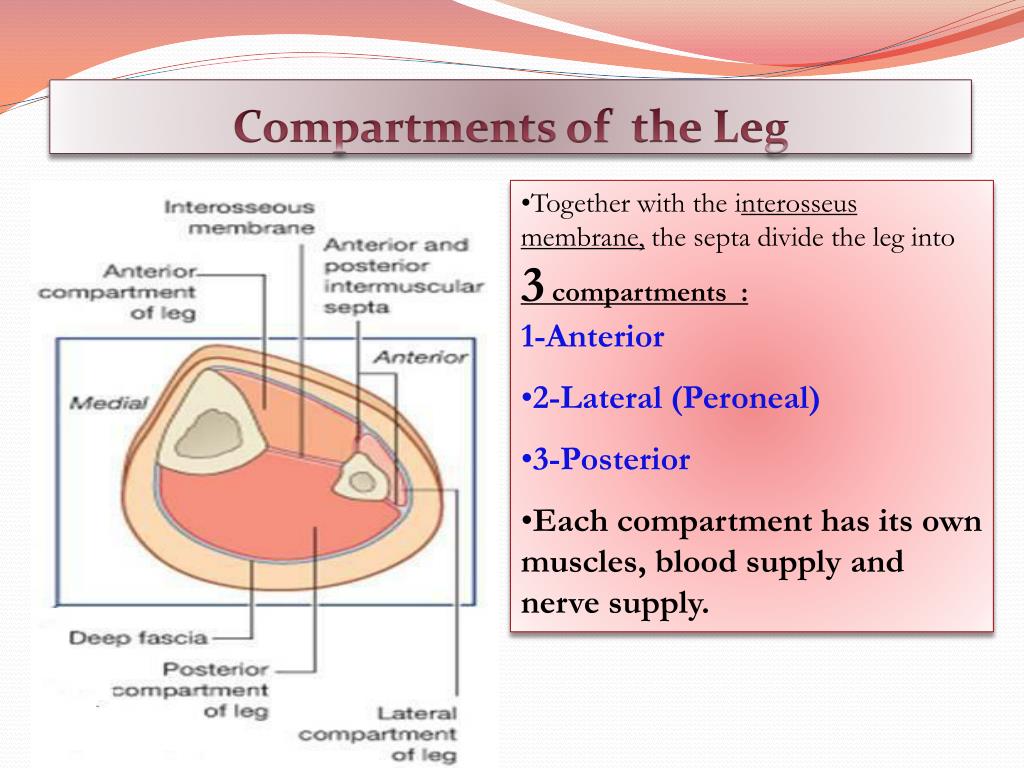

Fasciotomy can be performed either with classical open or minimally invasive techniques including endoscopically assisted or semi blind subcutaneous releases. Fasciotomy is currently the mainstay for surgical treatment. The nerve contains axons from the L4, L5, and S1 spinal nerves.īlood for the compartment is supplied by the anterior tibial artery, which runs between the tibialis anterior and extensor digitorum longus muscles. Compartment syndrome is a common cause of lower extremity pain via an increased intra-compartmental pressure. The anterior compartment of the leg is supplied by the deep fibular nerve (deep peroneal nerve), a branch of the common fibular nerve. When pressure is above diastolic blood pressure, arteries.

Inside each layer of fascia is a confined space, called. Pressure in the compartment builds and eventually compresses veins and nerves, reducing blood flow away from the muscles and sensation. Thick layers of tissue, called fascia, separate groups of muscles in the arms and legs from each other. Compartment syndrome occurs when there is a build-up of pressure inside a fascial compartment in a limb.

The compartment contains muscles that are dorsiflexors and participate in inversion and eversion of the foot. Clinical relevance: compartment syndrome 1. Inferior third of anterior surface of fibula and interosseous membraneĭorsiflexes ankle and aids in eversion of foot Middle and distal phalanges of lateral four digitsĮxtends lateral four digits and dorsiflexes ankle Lateral condyle of tibia and superior three quarters of medial surface of fibula and interosseous membrane in the distal half of the tibia the deep posterior compartment lies just below the subcutaneous tissue - again, releasing the fascia over the FDL is required. Middle part of anterior surface of fibula and interosseous membraneĭorsal aspect of base of distal phalanx of great toe (hallux) Fascial Compartments of Leg Leg: Cross Sections and Fascial Compartmen Fascial Compartments of Leg Leg: Cross Sections and Fascial Compartments Muscles: Cross. Studies examining fascial thickness and stiffness suggest that an overly noncompliant. This avoids unnecessary testing and, more importantly, subsequent referral for and even execution of surgical treatment ( 18,33). Medial and inferior surfaces of medial cuneiform and base of 1st metatarsal The lower leg contains four separate compartments, each bound by fascia and containing nerves and/or arteries and veins. Lateral condyle and superior half of lateral surface of tibia and interosseous membrane It contains muscles that produce dorsiflexion and participate in inversion and eversion of the foot, as well as vascular and nervous elements, including the anterior tibial artery and veins and the deep fibular nerve. Innervation: Superficial fibular (peroneal) nerve.The anterior compartment of the leg is a fascial compartment of the lower leg.Also supports the lateral and transverse arches of the foot. Actions: Eversion and plantarflexion of the foot.The tendon crosses under the foot, and attaches to the bones on the medial side, namely the medial cuneiform and base of metatarsal I.The fibres converge into a tendon, which descends into the foot, posterior to the lateral malleolus.

The fibularis longus originates from the superior and lateral surface of the fibula and the lateral tibial condyle. Lateral compartment: The lateral plantar fascia overlies the abductor and flexor digiti minimi brevis.The fibularis longus is the larger and more superficial muscle within the compartment. In reality, the job of these muscles is to 'fix' the medial margin of the foot during running, and prevent excessive inversion. Note: From the anatomical position, only a few degrees of eversion are possible. In this article, we shall look at the anatomy of the muscles in the lateral compartment of the leg - their attachments, innervation and actions. Chronic exertional compartment syndrome often occurs in the same compartment of an affected limb on both sides of the body, usually the lower leg. They are both innervated by the superficial fibular nerve. The common function of the muscles is eversion – turning the sole of the foot outwards. Step 1 single-incision technique position the patient: Place the patient supine with a bump underneath the ipsilateral buttock. There are two muscles in the lateral compartment of the leg the fibularis longus and brevis (also known as peroneal longus and brevis). Introduction: Compartment syndrome of the leg is an orthopaedic emergency and can be treated with single or dual-incision fasciotomy, allowing for necessary decompression of all four compartments.


 0 kommentar(er)
